| The
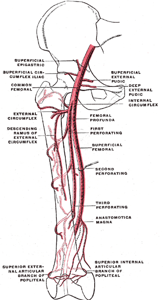
primary blood supply to the head of the femur arises from the
circumflex arteries located at the base of the femur neck.
Other arteries branch off from the circumflex arteries, and they
extend from the
femur neck towards the femur head. These arteries can become
disrupted by a hip fracture or hip dislocation, leading to
necrosis (i.e. bone death) of the femur head (i.e. avascular necrosis or
AVN).
The
principal sources of blood flow to the femoral head are vessels
branching off the medial femoral circumflex artery. They
run under the synovial membrane and enter
the femoral head within a 1-cm wide zone between the cartilage of
the femoral head and the cortical bone of the femoral neck.
They supply the lateral and central thirds of the femoral
head. The artery of the ligamentum teres--a secondary blood
source for the femur head-- supplies the medial third of the
femoral head.
The
branching arteries and the artery of the ligamentum teres
connect in the junction of the central and medial
third of the femoral head. The thickest part of the
articular cartilage of the femoral head is located along the
posterior-superior aspect and measures 3 mm in diameter. It thins
to 0.5 mm along the peripheral and inferior margins.
Trauma is the
most common cause of AVN (although there are other causes such
as steroid use and alcoholism), and it can occur within 8 hours
after a traumatic disruption of the blood supply. The
blood vessels can be damaged as
they enter the femur. The artery of the ligamentum teres
also may be damaged. In addition, intracapsular hematoma
can increase
intracapsular pressure, which can cause blockage of the vessels
within the joint capsule.
|

Image
from Gray's Anatomy |
|

Diagram
from research report by Alex Ying-Shyuan Lee, Howard Haw-Chang
Lan, San-Kan Lee on www.rsroc.org.tw |
Fractures of the
trochanter and those occurring outside the hip capsule rarely develop
AVN; but following hip dislocation, circulation is interrupted because of
tears of the ligamentum teres which can precipitate AVN. Tearing of
the joint capsule also compromises the vessels within the capsule. AVN
can develop as late as 10 years after fractures of the
femur.
Because of the
limited and delicate nature of the circulatory system feeding the
femoral head and neck, it is possible that the blood flow could
become disturbed due to the action of dislocating the hip and the
trauma of the surgery itself. The blood flow is disrupted
during resurfacing surgery, so a short surgical time is desirable.
Surgeons take great pains to
protect the surrounding arteries, but success is not
guaranteed. Therefore, resulting necrosis of the femur head
and neck remains one of the risk factors in hip resurfacing.
|

